This study aims to enhance our understanding of the complex mechanisms underlying visual impairment in DME patients, investigating large cysts as a potential biomarker for visual deterioration in DME patients.
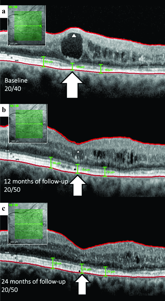
In the present study, we identified a correlation between large central macular intraretinal cysts and worse BCVA outcomes compared to their smaller counterparts, corroborating earlier studies [7]. We also observed a moderate correlation between CMT and cyst height with BCVA, which is consistent with earlier publications [3, 4, 9, 15,16,17]. Existing studies, including post hoc analyses of the RIDE/RISE and RESTORE/RESTORE-extension studies, support the hypothesis that an increase in cyst size may lead to poorer visual outcomes [18]. Large foveal cysts, mainly those greater than 380 μm, exhibit a significant deterioration in BCVA even after post-treatment reduction [4]. Figure 3 illustrates a case that supports the aforementioned statement and aligns with the results of our study.
Sequential evolution of a cyst in the inner nuclear layer (INL) and subsequent impingement on the outer retinal layer (ORL) in an included patient. a, Following the formation and growth phase, large cysts begin to compress the inner layers, as indicated by the arrowhead, which exhibits hyperreflectivity due to the compaction of cells and impinges on the ORL, as shown by the arrow. In this instance, the external limiting membrane (ELM), ellipsoid zone (EZ), and interdigitation zone (IZ) are thinned and appear to disappear. The ORL thickness measured at different points – underneath the cyst and at 500 microns temporal and nasal – were 60, 80, and 98 microns, respectively. b, Over the one-year follow-up under bevacizumab treatment, the cyst height decreased, allowing the ellipsoid zone to be identifiable once more, albeit with disruption (indicated by a black asterisk). Concurrent ORL thinning is also observed. The ORL thickness was measured at different points: 72, 79, and 96 microns underneath the cyst and 500 microns both temporally and nasally. c, Ultimately, there is noticeable disruption of the ellipsoid zone and ORL thinning after the resolution of the cyst, without concurrent retinal pigment epithelium (RPE) atrophy in this case, as marked by the black asterisk
When evaluating the CMT, we observed that it is composed of cysts of various sizes. Smaller intraretinal cysts located at the foveal center have been associated with better visual outcomes compared to larger cysts, as noted by Gerendas and Pelosini et al. [4, 9] Thus, when CMT is predominantly composed of smaller cysts, visual function tends to be less affected than when larger cysts are present. This variability in cyst size may help explain the low to moderate correlation between CMT and BCVA reported in multiple studies. Our findings suggest that there may be a threshold in cyst growth, beyond which retinal neurons are at greater risk of damage. This emphasizes the critical need for timely intervention in the management of DME and highlights the importance of early treatment to preserve visual function.
We also explored the evolving morphology of enlarging cysts, whereupon reaching a critical volume – constrained by the retina’s accommodative elasticity – they begin to spread laterally, changing their characteristic spherical form to a posterior flat conformation, i.e., the cyst plateau. This transformation may be influenced by the relative stiffness of the ORL, leading to photoreceptor compression. Indeed, when evaluating the general trend of the degree of elasticity across the various retinal layers, a gradual increase in rigidity is observed towards the outermost layers, with the photoreceptor layer being the least elastic [19, 20]. This phenomenon could explain the significant correlation observed in our study between the cyst plateau and worse BCVA, as well as a higher incidence of EZ disruption.
In a groundbreaking study, Karahan et al. reported that cysts within the retina can exert substantial pressure on the internal retinal layers, potentially leading to significant damage. They developed a mathematical formula that demonstrated that, in some scenarios, the cyst could generate a pressure of approximately 60 mmHg, potentially impeding axoplasmic flow and causing injury to the retinal nerve fiber layers [10]. However, their model proposed that the pressure exerted by the intraretinal cystoid space was equivalent to the force applied per unit area, a simplification that may not fully capture the complexity of the process involved. Although our study did not investigate the effects of cysts on the inner retinal layers, we propose an additional explanation through the mechanical compression of cysts on the ORL to rationalize the more deleterious impact on vision observed with large cysts compared to smaller ones.
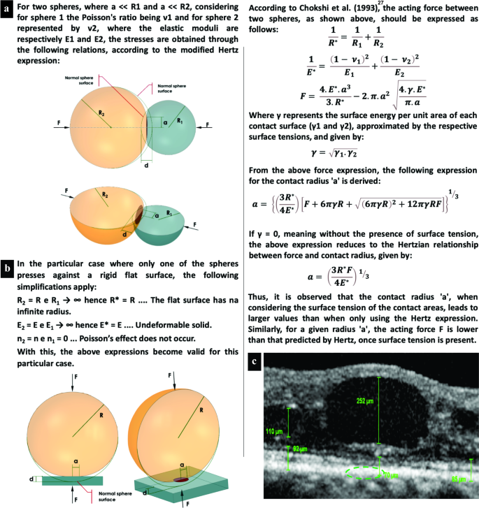
To further explore this phenomenon, we suggest applying Hertz’s theory of contact mechanics [12]. This theory can be used to measure the tension in the contact area, depending on the applied forces, the radii of curvature of the two bodies, and their moduli of elasticity. It is also possible to calculate the size of the contact area and the depth of deformation of the two bodies, represented by simple geometric figures, such as two spheres or a sphere and a flat surface (Fig. 4a and b).
Schematic representation of contact stress and deformation according to contact mechanics. a, The image illustrates two interacting spheres where the point of maximum contact pressure is located centrally, resulting in a semi elliptic pressure distribution, as depicted by the green dashed elliptic area in C. b and c, These images demonstrate that the theory is applicable even when a spheroid object, such as a retinal cyst, engages with a more or less flat surface, akin to the retina. This principle holds because even a “flat” plate can be equated to a sphere with an infinite radius, as shown in B
Legends: F, forces between the bodies; R, radii of curvature; a, radius of the contact area; d, depth of deformation; E, moduli of elasticity; ν, Poisson’s ratios; d, depth of identation; R*, flat surface with an infinite radius; E*, undeformable solid
According to Hertz´s theory, the maximum contact pressure arises at the center of the interacting spheres, giving rise to a semi elliptic pressure distribution. Theoretically, the contact area of two spheres is a single point. As a result, high pressure, which tends to infinity, arises between the two curved surfaces, causing immediate deformation of both surfaces. However, owing to the elastic deformation of the bodies, the contact point is converted into a small contact area [12, 21]. We believe that this theory can be useful in understanding the development of a posterior flat conformation in large cysts when certain critical volumes are reached according to the accommodative elasticity of the retina. Nevertheless, the classical Hertz model has limitations since it remains accurate only when the ratio of the indentation depth to the indenter radius is less than 0.1 [22].
In real-world scenarios, materials with different properties interact, leading to a diverse range of indentations. This has spurred several authors to evolve the Hertz model to characterize contact behaviors under finite indentations using both linear and nonlinear regimes of elasticity [23,24,25,26,27], akin to the approach we have adopted in our study. These refined models (modified theories), validated through comparative simulations with the classical Hertz theory, revealed congruent curve patterns, thereby substantiating the applicability of the fundamental principles of the theory.
Furthermore, when applying the modified Hertz theory in scenarios involving liquids, it is imperative to account for surface tension — a critical factor highlighted in Chokshi’s 1993 publication. The article ingeniously incorporated this aspect into Hertzian theory, underscoring its practicality in elucidating the microphysics of coagulation between two colliding, smooth, spherical grains at the elastic limit and demonstrating its adaptability to analyses involving diverse materials or tissues [28].
In our clinical observations, we noted that large cysts exhibit signs of compressing the ORL, resulting in its thinning (Fig. 4c) and adopting a flat punch shape of pressure distribution [29]. This phenomenon can be conceptualized through Hertzian theory, which outlines how perpendicular and tangential forces interact between spheres at the point of contact (Fig. 4a). Intriguingly, this theory retains its applicability even when a spherical entity, such as a retinal cyst, contacts a substantially flat surface, such as the ORL (Fig. 4c). This is attributable to the theoretical equivalence of a flat plate to a sphere with an infinite radius (Fig. 4b).
In alignment with the assumption that a retinal cyst functions analogously to a sphere, our investigation suggests the presence of both perpendicular and tangential contact forces at the junction between the cyst and the ORL. Furthermore, as the cyst enlarges, increasing its contact with the ORL, reciprocal deformation occurs, characterized by plateauing and compression phenomena, respectively (Fig. 1.2b, Fig. 3a, and Fig. 4c) [11].
In our cohort, ORL thinning was observed exclusively in eyes with a plateau, and this thinning was correlated with both worse BCVA and increased cyst height according to multivariate regression analysis. Despite the absence of a direct correlation between ORLt and BCVA, a greater number of eyes with ORL thinning exhibited a decline in BCVA. We hypothesize that the minor variations in ORLt, combined with the sample size, might be the underlying factors preventing the achievement of statistical significance.
In light of the findings gained from this research, we acknowledge the suitability of the modified Hertz’s theory of contact mechanics as a congruent model to elucidate our observations. This theoretical framework provides a more profound understanding of the adverse effects that large intraretinal cysts exert on the retina and vision, thereby reinforcing its crucial role as a pivotal biomarker in diabetic macular edema (DME).
Our findings, while clinically significant, should be approached with caution due to several limitations inherent to this study. There is potential for other underlying structural alterations induced by DR concurrent with macular edema; these alterations can affect the integrity of both the ORL and the inner retinal layers, leading to damage to other cells, including bipolar cells, thereby contributing to visual dysfunction. Moreover, the relatively small sample size constitutes a limitation, as the compressed inner retinal layers situated above the cyst were not evaluated for thickness, nor was the foveal avascular zone measured. Although linear rather than volumetric measurements were taken during the analysis of the morphological characteristics of the cysts, the results obtained provide practicality for clinical use. Given these constraints, we recommend interpreting the results carefully while acknowledging the need for further research to substantiate our findings with a more comprehensive analysis encompassing a larger and more diverse patient cohort.


Leave a Reply